Subacute cutaneous lupus erythematosus
Skin lesions of subacute cutaneous lupus erythematosus (SCLE) are annular or psoriasiform. There is no follicular plugging, no scarring, and little atrophy, all of which are frequently noted in CCLE. Patients with SCLE may have a few of the criteria of SLE as defined by the American Rheumatism Association, including photosensitivity, arthralgias, serositis, renal disease, and serologic abnormalities; practically all have anti-Ro (SS-A) and most have anti-La (SS-B) antibodies. The serious criteria of SLE are uncommon: severe vasculitis, severe CNS disease, or progressive renal disease. Nonetheless, most patients have a mild form of SLE so that SCLE is not a purely cutaneous disease. The clinical skin lesions are the distinctive feature of SCLE.
Causes of Cutaneous Lupus Erythematosus
Physical examination
Skin lesions Two Types Psoriasiform Papulosquamous, sharply defined, with slight delicate scaling, evolving into bright red confluent plaques that are oval, arciform, or polycyclic, just as in psoriasis.
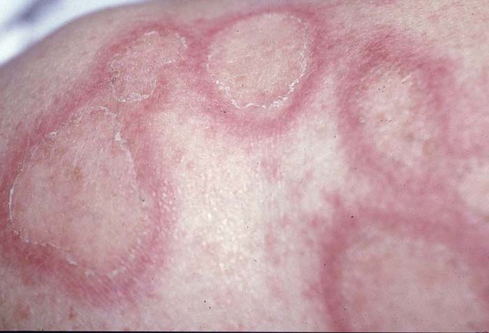
Annular Bright red annular lesions with central regression and little scaling. In both there may be telangiectasia, but there is no follicular plugging and less induration than in CCLE. Lesions resolve with slight atrophy (no scarring) and hypopigmentation.
Other Lesions Periungual telangiectasia, diffuse nonscarring alopecia.
Distribution Scattered, disseminated in lightexposed areas: shoulders, extensor surface of the arms, dorsal surface of the hands, upper back, V-neck area of the upper chest.
Diagnosis
Clinical findings confirmed by histology and immunopathology. The extensive involvement is far more than is ever seen in CCLE, and the distinctive eruption is a marker for SCLE.
Treatment
Topical Anti-inflammatory glucocorticoids are only partially helpful.
Chronic cutaneous lupus erythematosus (CCLE)
This chronic, indolent skin disease is characterized by sharply marginated, scaly, infiltrated, and later atrophic red (“discoid”) plaques, usually occurring on habitually exposed areas. This disorder, in most cases, is purely cutaneous without systemic involvement. However, CCLE lesions may occur in SLE. CCLE may manifest as chronic discoid LE (CDLE) or LE panniculitis.
Classic chronic discoid LE(CCLE)
History
Can be precipitated by sunlight but to a lesser extent than ACLE or SCLE. Lesions last for months to years. Usually no symptoms, some times slightly pruritic or smarting. No general symptoms.
Physical examination
Skin lesions Bright red papules evolving into plaques, sharply marginated, with adherent scaling. Scales are difficult to remove and show spines on the undersurface (magnifying lens) resembling carpet tacks. Plaques are round or oval, annular or polycyclic, with irregular borders and expand in the periphery and regress in the center, resulting in depression of lesions, atrophy, and eventually scarring. Follicular plugging (closely set and often in clusters) and dilated follicles may persist in atrophic and scarred lesions but eventually disappear so that smooth, whitish scarred lesions result that are partially surrounded by a still active inflammatory and raised border. While active lesions are bright red, “burned out” lesions may be pink or white (hypomelanosis) macules and scars, but scarred lesions may also show hyperpigmentation, especially in persons with brown or black skin.
Distribution and sites of predilection CDLE may be localized or generalized, occurring predominantly on the face and scalp; otherwise: dorsa of forearms, hands, fingers, toes, and less frequently, the trunk.
Scalp Scarring alopecia with residual inflammation and follicular plugging.
Mucous Membranes Less than 5% of patients have lip involvement (hyperkeratosis, hypermelanotic scarring, erythema) and atrophic erythematous or whitish areas with or without ulceration on the buccal mucosa, tongue, and palate.
Diagnosis
Clinical findings confirmed by histology.
Treatment
Prevention Topical sunscreens (SPF 30) routinely.
Local Glucocorticoids Topical fluorinated glucocorticoids (with caution). Intralesional triamcinolone acetonide, 3 to 5 mg/ml, for small lesions.
Antimalarial Hydroxychloroquine, ≤6.5 mg/kg of body weight per day. If hydroxychloroquine is ineffective, add quinacrine, 100 mg tid. Monitor for side effects.
Retinoid Hyperkeratotic CDLE lesions respond well to systemic etretinate (1 mg/kg of body weight).
References