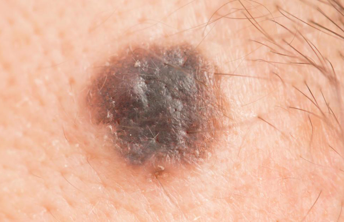
Nodular melanoma (NM) is second in frequency (14%) after superficial spreading melanoma (SSM) in most series, occurring largely in middle life in persons with white skin and, as in SSM, on the less commonly exposed areas. The tumor from the beginning is in the “vertical growth” phase and could appropriately be called a deeply penetrating melanoma (as opposed to SSM). NM is uniformly elevated and presents as a thick plaque or an exophytic, polypoid or dome-shaped lesion. The color pattern is usually not variegated, and the lesion is uniformly blue or blue-black or, less commonly, can be very lightly pigmented or nonpigmented (amelanotic melanoma) and confused with a pyogenic granuloma or other nonpigmented tumors. NM is the one type of primary melanoma that arises quite rapidly (4 months to 2 years) from normal skin or from a melanotic nevus as a nodular (vertical) growth without an adjacent epidermal component, as is always present in SSM and LMM.
Causes of Nodular Melanoma
Both SSM and NM occur in approximately the same sites (upper back in males, lower legs in females), and presumably the same pathogenetic factors are operating in NM as were described in SSM. For the growth pattern of NM. The reason for the high frequency of NM in the Japanese is not known.
Symptoms of Nodular Melanoma
NM usually occurs on sun-exposed areas like the head (scalp in men), neck, trunk, arms and legs but can arise anywhere. It is more frequent in males than females, most common during middle age, and generally doesn’t grow from an existing mole starting instead as a new growth of its own.
Growth is rapid both below and above the skin frequently achieving a visible diameter of 1-2 cm or larger. Because of this, they are easy to notice, often resembling a blood blister. Unfortunately, most people wait an average of nine months to have a lesion checked. NM grows in weeks to months, not months to years.
Diagnosis
- Skin biopsy
- Regular self-examinations
- The dermoscopic appearance is particularly helpful in the diagnosis of early melanoma, but in general only specialist dermatologists are trained in dermoscopy and dermoscopy is not necessary if the lesion has the typical clinical appearance of melanoma.
Treatment
Biopsy Total excisional biopsy with narrow margins-optimal biopsy procedure, where possible. Incisional or punch biopsy acceptable when total excisional biopsy cannot be performed or when lesion is large, requiring extensive surgery to remove the entire lesion.
Surgical Treatment
Dermatopathology Malignant melanocytes, which appear as epitheloid, spindle, or small atypical cells, show little lateral (radial) growth within and below the epidermis and invade vertically into the dermis and underlying subcutaneous fat. They are S-100 and usually HMB-45 positive.
- https://www.dermnetnz.org/topics/nodular-melanoma/
- https://www.ncbi.nlm.nih.gov/pmc/articles/PMC2644319/