Superficial phlebitis is an inflammatory thrombosis of a superficial normal vein, usually due to infection or trauma from needles and catheters, or of a varicose vein usually in the context of the chronic venous insufficiency (CVI) syndrome. Deep venous thrombosis is due to thrombotic obstruction of a vein with or without an inflammatory response and occurs due to slow blood flow, hypercoagulability, or changes in the venous walls.
Causes of Thrombophlebitis and Deep Venous Thrombosis
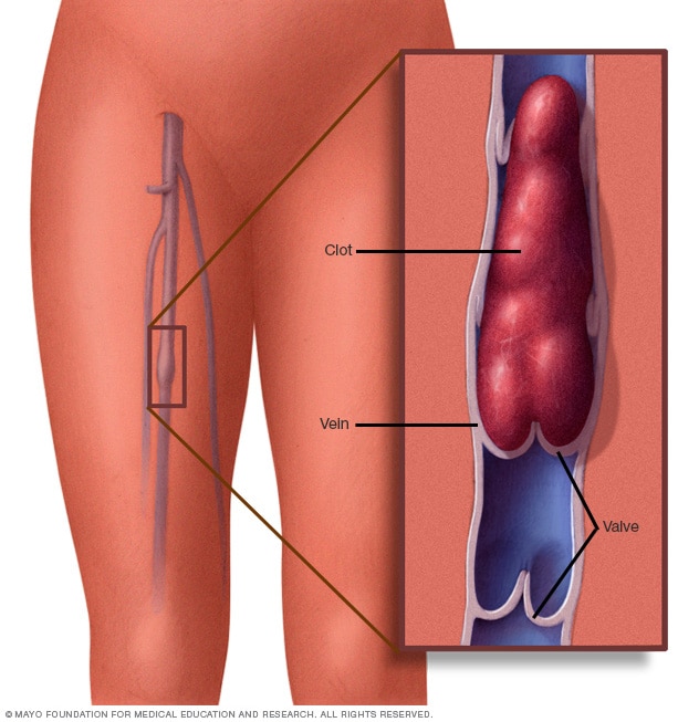
The thrombus originates in an area of low venous flow. An occlusion of a vein by thrombus imposes a block to venous return which leads to increased venous pressure and edema in the distal limb. An inflammatory response to the thrombus causes pain and tenderness. If the venous pressure is too high, arterial limb flow may rarely be compromised and ischemia of the distal limb may occur. The thrombus in the vein often has a free-floating tail, which may break off to produce a pulmonary embolus. Organization of the thrombus in the vein destroys the venous walls, and this leads to post-phlebitic syndrome.
Symptoms of Thrombophlebitis and Deep Venous Thrombosis
- Swelling
- Gradual onset of pain
- Redness
- Warm to touch
- Worsening leg pain when bending the foot
- Leg cramps, especially at night
- Bluish or whitish discoloration of skin
- pain, or an aching heavy sensation in the limb, sudden swelling, tenderness, or discoloration of the limb
- slight fever
Diagnosis
Diagnosis based on clinical exam alone can be difficult since none of the symptoms of thrombophlebitis or DVT are unique and can be due to other disorders. Objective testing is mandatory to establish the diagnosis. There is no specific blood test available that readily identifies a diagnosis of DVT.
Duplex ultrasonography is a noninvasive test that is the preferred initial study for DVT. It is a combination of real-time B-mode ultrasound and Doppler assessment. An ultrasound beam is used to locate areas of vein obstruction that are indicative of a thrombus.
Contrast venography is a procedure that is used when other less invasive tests are inconclusive. It is the only reliable test for patients who are asymptomatic (without symptoms). Dye is injected into a vein in the foot. The dye allows visualization of the leg veins to determine whether there are areas of defect that indicate a blockage of the vein by a thrombus.
Impedance plethysmography is a noninvasive method in which a pneumatic cuff is placed around the thigh and inflated. A mild electrical current is passed through the leg and provides information on blood flow when the cuff is deflated.
Treatment
The treatment of deep venous thrombosis is anticoagulation. IV heparin is given at a loading dose of 5000 U and approximately 1000 U/h thereafter. The partial thromboplastin time (PTT) should be 1.5 to 2 times normal. Low-molecular-weight heparin is also effective, and warfarin can be started orally at the same time and should overlap heparin for 5 days until the necessary factors for blood clotting are depressed. Patients should be treated for 3 months with anticoagulation. Elastic stockings and compression are mandatory and should be worn for at least three months, and ambulation should be started as soon as symptoms subside.
References
- https://www.ncbi.nlm.nih.gov/pubmed/9290540
- https://www.unmc.edu/surgery/divisions/gensurg/vascular/patient-care/diseases/dvt.html
- https://www.empr.com/patient-fact-sheets/deep-vein-thrombosis-dvt-and-thrombophlebitis-patient-information-fact-sheet/article/219839/