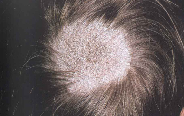
Tinea capitis is a dermatophytic trichomycosis of the scalp. Clinical presentations vary widely, ranging from mild scaling and broken-off hairs to severe, painful inflammation with painful, boggy nodules that drain pus and result in scarring alopecia.
Causes of Tinea Capitis
Noninflammatory lesions Invasion of hair shaft by the dermatophytes, principally M.audouinii (child-to-child, via barber, hats, theater seats), M. canis (young pets-to-child and then child-to-child) or T.tonsurans. Inflammatory lesions: T tonsurans, M.canis, T.verrucosum, and others. Spores enter through breaks in hair shaft or scalp to cause clinical infection.
Scalp hair traps fungi from the environment or fomites. Asymptomatic colonization is common Trauma assists inoculation. Dermatophytes initially invade stratum corneum of scalp, which may be followed by hair shaft infection. Spread to other hair follicles then occurs. Eventually, infection regresses with or without an inflammatory response. Clinical appearance varies with type of hair invasion, level of host resistance, degree of inflammatory host response: few dull-grey, broken-off hairs with little scaling to severe painful inflammatory mass covering entire scalp. Partial hair loss with inflammation in all cases. Kerion is associated with a high degree of hypersensitivity to fungal hapten. Types of hair invasion:
Microsporum types:
- Small-spored ectothrix; hair shaft is invaded in mid-follicle. Intrapiliary hyphae grow inward toward hair bulb. Secondary extrapiliary hyphae burst, growing over surface of hair shaft.
- Large-spored ectothrix have similar arrangement.
Trichophyton types:
- Large-spored ectothrix (in chains); arthrospores spherical, arranged in straight chains, confined to external surface of hair shaft. Spores are all larger than those of small-spored Microsporum ectothrix.
- Entothrix type; intrapiliary hyphae fragment into arthroconidia within hair shaft making it fragile, with subsequent breakage close to scalp surface.
Symptoms of Tinea Capitis
- Itching of the scalp, may be slight or absent
- Skin (scalp) lesions that are: Round, scaly, Gray or reddened (skin redness or inflammation), Bald-appearing patches (hair is broken off, not actually missing), Possibly small black dots on the scalp
- Occasionally localized area of swelling, raw skin, or pus-filled lesion on the scalp (kerion)
Diagnosis
Doctors can diagnose ringworm on sight, or they may take a skin scraping. This is examined under a microscope, or put on an agar plate in a microbiology laboratory and allowed to grow. Some of the fungi fluoresce under a black light examination.
Treatment
Topical antifungal agents Topical agents are ineffective in management of tinea capitis. Duration of treatment should be extended until symptoms have resolved and fungal cultures negative.
Oral antifungal agents Of the systemic antifungals available, terbinafine and itraconazole are superior to ketoconazole and all three to griseofulvin. Side effects in increasing order: terbinafine<itraconazole<ketoconazole< griseofulvin.
- Terbinafine 250 mg qd. Reduce dosing according to weight in pediatric patients.
- Itraconazole 100-mg capsules or oral solution (10 mg/mL). Treatment duration: 4 to 8 weeks. Pediatric Dose 5 mg/kg/d, Adult Dose 200 mg/d
Adjunctive therapy
- Prednisone 1 mg/kg/d for 14 days for children with severe, painful kerion.
- Systemic antibiotics For secondary S. aureus or group A Streptococcus infection, erythromycin, dicloxacillin, or cephalexin
Surgery Drain pus from kerion lesions.
Prevention
Important to examine home and school contacts of affected children for asymptomatic carriers and mild cases of tinea capitis. Ketoconazole or selenium sulfide shampoo may be helpful in eradicating the asymptomatic carrier state.
References